Hand Surgery Source

- Scaphoid Fracture -

You must be logged in to view this content.


Please Log In or Create Your Free Account.

FRACTURE, SCAPHOID

Introduction

Fracture Nomenclature for Scaphoid Fractures and Fracture Dislocations

Hand Surgery Resource’s Diagnostic Guides describe fractures by the anatomical name of the fractured bone and then characterize the fracture by the Acronym:

In addition, anatomically named fractures are often also identified by specific eponyms or other special features.

For the Scaphoid Fractures and Fracture Dislocations, the historical and specifically named fractures include:

Scaphocapitate fracture syndrome

Scaphoid dislocation and fracture-dislocation

Scaphoid avulsion fracture

Trans-scaphoid perilunate dislocation and fracture-dislocation

By selecting the name (diagnosis), you will be linked to the introduction section of this Diagnostic Guide dedicated to the selected fracture eponym.


Fractures of the carpal bones account for ~6% of fractures overall and up to 18% of all hand fractures. The vast majority (58-89%) occur in the scaphoid, while fracture of the other 7 carpals is very uncommon and only comprise ~1.1% of all fractures. Scaphoid fractures represent 2-7% of all orthopedic fractures and are second only to the distal radius in terms of frequent wrist injuries. They are most often seen in young men and the typical injury mechanism is forced wrist dorsiflexion, often from a fall on an outstretched hand (FOOSH), although direct blows and axial loading may also be responsible. Treatment for scaphoid fractures is controversial and preferences vary between treatment centers, but it is generally agreed that conservative treatment with cast immobilization is appropriate for nondisplaced and minimally displaced fractures, especially those in the distal third. Surgery is usually required for most proximal pole fractures, nondisplaced or unstable fractures, and cases that fail conservative interventions, as well as for nonunion after treatment, which is common. Prompt diagnosis and suspicion of scaphoid fractures can help increase the chances of a successful outcome and reduce the risk of complications.1-7

Definitions

  • A scaphoid fracture is a disruption of the mechanical integrity of the scaphoid.
  • A scaphoid fracture produces a discontinuity in the scaphoid contours that can be complete or incomplete.
  • A scaphoid fracture is caused by a direct force that exceeds the breaking point of the bone.

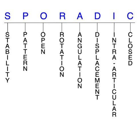
Hand Surgery Resource’s Fracture Description and Characterization Acronym

SPORADIC

S – Stability; P – Pattern; O – Open; R – Rotation; A – Angulation; D – Displacement; I – Intra-articular; C – Closed


S - Stability (stable or unstable)

  • Universally accepted definitions of clinical fracture stability are not well defined in the hand surgery literature.8-10
  • Stable: fracture fragment pattern is generally nondisplaced or minimally displaced. It does not require reduction, and the fracture fragment’s alignment is maintained with simple splinting. However, most definitions define a stable fracture as one that will maintain anatomical alignment after a simple closed reduction and splinting. Some authors add that stable fractures remain aligned, even when adjacent joints are put to a partial range of motion (ROM).
  • Unstable: will not remain anatomically or nearly anatomically aligned after a successful closed reduction and simple splinting. Typically unstable scaphoid fractures have significant deformity with comminution, displacement, angulation, and/or shortening.
  • The most commonly used classification system for scaphoid fractures is the Herbert classification, which is helpful because it specifically defines stable and unstable fractures.
    • Type A: stable
      • Type A1: fracture of tubercle
      • Type A2: incomplete fracture through waist
    • Type B: unstable
      • Type B1: distal oblique fracture
      • Type B2: complete fracture of waist
      • Type B3: proximal pole fracture
      • Type B4: trans-scaphoid perilunate fracture-dislocation
      • Type B5: comminuted fracture
    • Type C: delayed union
    • Type D: established non-union
      • Type D1: fibrous union
      • Type D2: pseudoarthrosis
      • Type D3: sclerotic pseudoarthrosis
      • Type D4: avascular necrosis11
  • In the Russe classification system, scaphoid fractures are more simply categorized as horizontal oblique, vertical oblique, or transverse. Under to this system, vertical oblique fractures only account for 5% of scaphoid fractures but result in the most shear forces across the fracture site, making it the least stable of these fracture types.3,12

P - Pattern

  • Scaphoid tubercle: only account for ~8% of scaphoid fractures.13
    • Caused by direct impact with the hand in dorsiflexion, ulnar deviation, and pronation, leading to a fragment avulsion by the radial collateral ligament, capsular attachments, or transverse carpal ligament14
  • Scaphoid distal pole: usually oblique
    • Account for ~10% of scaphoid fractures13
  • Scaphoid waist: transverse, vertical oblique, horizontal oblique, coronal, and/or comminuted; can be complete or incomplete
    • The most common fracture site, accounting for ~65% of all scaphoid fractures.13
    • Compressive forces are seen at the fracture site in horizontal oblique fractures, while transverse fractures have a combination of shear and compressive forces.3
    • Coronal fractures are rare and often missed and may be either complete and involve the entire waist, or partial and involve either the proximal or distal pole.15
  • Scaphoid proximal pole: unstable fractures that are difficult to treat2
    • May be easily missed on plain radiographs.3
    • Account for ~15% of scaphoid fractures.13
  • The usual mechanism of injury in scaphoid fractures is a forced dorsiflexion wrist injury—most commonly a FOOSH with a pronated and ulnarly deviated hand, and the wrist typically in wrist hyperextension past 95°—which causes longitudinal loading of the scaphoid and a subsequent fracture as the volar cortex fails in tension. The force then extends to the dorsal cortex, which fails in compression. Other possible injury mechanisms include a direct blow or axial loading with the wrist in neutral flexion-extension.2-4

O - Open

  • Open: a wound connects the external environment to the fracture site. The wound provides a pathway for bacteria to reach and infect the fracture site. As a result, there is always a risk for chronic osteomyelitis. Therefore, open fractures of the scaphoid require antibiotics with surgical irrigation and wound debridement.8,16,17
  • Open fractures of the scaphoid may require surgical exploration to determine if articular surfaces are involved. After irrigation and debridement, these wounds are generally left open and further treatment is typically delayed until the wound shows no sign of infection.18,19

R - Rotation

  • Scaphoid fracture deformity can be caused by rotation of the distal fragment on the proximal fragment.
  • Degree of malrotation of the fracture fragments can be used to describe the fracture deformity.
  • Scaphocapitate fracture syndrome consists of concomitant fractures of the scaphoid and capitate with a rotation of 90-180° of the proximal fragment of the capitate.20
  • Opposing rotational moments on the proximal and distal poles cause dorsal angulation in scaphoid fractures.4
  • The violent dorsiflexion force involved in most scaphoid often causes the scaphoid to rotate out of its correct anatomical position while maintaining the integrity of the ligaments supporting the lunate, triquetrum, and hamate.21
  • With a fracture to its waist, the distal scaphoid rotates around the radioscaphocapitate ligament into an abnormal flexion, ulnar deviation, and pronation posture, while the lunate and attached proximal part of the scaphoid rotate into an abnormal extension, supination, and radial deviation.22

A - Angulation (fracture fragments in relationship to one another)

  • Angulation is measured in degrees after identifying the direction of the apex of the angulation.
  • Straight: no angulatory deformity
  • Angulated: bent at the fracture site
  • Failure to recognize a scaphoid fracture may result in humpback deformity of the scaphoid and dorsal angulation of the lunate—or dorsal intercalated segmental instability (DISI) deformity—which is caused by opposing rotational moments on the proximal and distal scaphoid poles.3,4
  • Some authors have defined displacement in scaphoid fractures as a gap of 1 mm on plain radiographs, scapholunate angle of >60°, or a radiolunate angle of >15°.3

D - Displacement (Contour)

  • Displaced: disrupted cortical contours
  • Nondisplaced: fracture line defining one or several fracture fragments; however, the external cortical contours are not significantly disrupted
  • The majority of scaphoid fractures are stable and nondisplaced.
  • Fracture displacement of >1 mm is associated with instability and is an indication for surgical intervention—such as open reduction and internal fixation (ORIF)—since these fractures heal more slowly, require longer immobilization, and are associated with a higher risk of non-union and post-traumatic osteoarthritis.4,7
  • Vertical oblique scaphoid fractures are more likely to be displaced by shear forces, while horizontal oblique and transverse fractures have greater compressive forces and are less likely to be displaced.23,24

I - Intra-articular involvement

  • Fractures that enter a joint with one or more of their fracture lines.
  • Scaphoid fractures can have fragment involvement with the radius or any of its intercarpal joint articulations.
  • If a fracture line enters a joint but does not displace the articular surface of the joint, then it is unlikely that this fracture will predispose to post-traumatic osteoarthritis. If the articular surface is separated or there is a step-off in the articular surface, then the congruity of the joint will be compromised, and the risk of post-traumatic osteoarthritis increases significantly.
  • Scaphoid fractures have a tendency for nonunion due to the nature of intra-articular fractures and the vulnerable vascularity of the proximal fragments.7

C - Closed

  • Closed: no associated wounds; the external environment has no connection to the fracture site or any of the fracture fragments.8-10
  • Whether a trans-scaphoid perilunate fracture-dislocation is open or closed is one of the major factors that will help determine its clinical outcome.25

Scaphoid fractures: named fractures, fractures with eponyms and other special fractures

Scaphocapitate fracture syndrome

  • Rare but complex injury that is typically considered a manifestation of the perilunate injury pattern.1,26
    • Some believe this injury represents the final stage of a greater arc injury, but the different degrees and directions of displacement of the capitate head imply that it does not have a unique mechanism.20,26,27
  • Most commonly occurs in young men between ages 20-30.28
  • The most recognized mechanism of injury is a volar-applied force to a hyperextended wrist, such as from a fall from a height or vehicular accident.28
    • This wrist hyperextension results in a scaphoid fracture and, with further extension, the capitate impacts on the dorsal lip of the radius. This produces a transverse capitate body fracture, in which its proximal fragment rotates 90-180° in the sagittal plane as the hand returns to a neutral position.1
  • Can be either isolated or associated with a perilunate dislocation, but a substantial force is usually required to cause a dislocation.28
  • In one study, scaphocapitate fractures were classified into the following 6 types based on fragment geometry and displacement:
    • Type I: transverse fracture of the scaphoid and capitate without dislocation
    • Type II: inverted proximal fragment of capitate that remains in articulation with the lunate
    • Type III: dorsal perilunate dislocation
    • Type IV: volar perilunate dislocation of the carpus and proximal fragment of the capitate
    • Type V: isolated volar dislocation of the proximal fragment of the capitate
    • Type VI: isolated dorsal dislocation of the proximal fragment of the capitate26

Imaging

  • Plain radiographs may not show the extent of the median nerve lesion. This, combined with the rarity and complexity of these injuries, causes many diagnoses to be initially missed or incorrectly labelled as simple scaphoid fractures.20,26
  • If plain radiography does not lead to a satisfactory diagnosis, a CT scan may be needed, especially if a complex carpal lesion is suspected.20,26

Treatment

  • Some controversy exists about the optimal management strategy for these injuries, but it appears that conservative treatment—consisting of closed reduction and cast immobilization—may be appropriate for some nondisplaced scaphoid and capitate fractures.26
  • For displaced or comminuted fractures, ORIF is generally considered the treatment-of-choice to reduce complications. The aim of treatment is reduction and fixation of both fractures to obtain bony union.
    • A dorsal approach is most commonly used, while a volar approach is usually reserved for when decompression of the median nerve is necessary.22
    • K-wires or compression screws are typically recommended to achieve fixation and reduce the risk for nonunion, and no significant differences have been identified between these approaches.20,28
  • According to the literature, it is recommended that reduction of the capitate precedes reduction of the scaphoid.
    • Reduction of the scaphoid fragments is guided from the radial surface of the capitate and is not easily maintained if the capitate is not stabilized, as the proximal scaphoid fragment tends to displace into the gap of the capitate head.20
  • Some authors recommended excision of the displaced proximal capitate fragment because of the possibility of avascular necrosis, but it’s been found that the capitate head can revascularize when replaced anatomically and immobilized until fracture healing.21
    • Excision can also interfere with the function of the carpus and eventually result in osteoarthritis. It is therefore advised that the capitate fragment is not excised, even if it cannot be fixed.26
  • Cancellous bone grafting may be considered in some cases of bone loss.20

Complications

  • Infection
  • Avascular necrosis
  • Non-union
  • Osteonecrosis

Outcomes

  • Early surgical intervention with meticulous reduction and fixation of all fractures and dislocations present generally leads to a favorable result with minimal complications. Despite the severity of this injury, restoring normal anatomical relationships of the carpus can lead to a successful long-term functional outcome.20
  • In one study of individuals with nondisplaced scaphoid fractures, those treated surgically returned to work earlier and had better functional outcomes than those treated conservatively.29
  • Delayed diagnosis and treatment of this injury has been found to result in non-union with subsequent carpal arthritis and carpal collapse.26

Scaphoid dislocation and fracture-dislocation

  • Scaphoid dislocation represents a wide spectrum of injuries with varying extent of ligamentous damage and may occur either in isolation or concomitantly with fracture(s) or other dislocation(s).
    • In general, these injuries are rare, but isolated scaphoid dislocation without another associated fracture or dislocation is even more uncommon. This is because a substantial amount of force is required to disrupt the scaphoid from its fossa, and when this occurs, the radial styloid or scaphoid waist are typically also fractured.21,23,30
    • A recent classification system has grouped isolated scaphoid dislocations into the following categories: primary or secondary, simple or complex, and partial or total.
      • Primary dislocations result directly from the injury, while secondary dislocations persist after failed closed reduction.
      • Simple dislocations involve only the scapholunate and radioscaphoid, while complex types have distal carpal row involvement.
      • In partial dislocations, the proximal pole is found outside the fossa, but the soft tissue attachments to the distal portion are intact. Complete dislocations refer to injuries in which the dislocated scaphoid lacks any soft tissue attachments.23,30
      •  
  • The exact mechanism of injury is not known, but it is generally believed that scaphoid dislocations are caused by violent dorsiflexion of the wrist with the hand grasping a fixed object in ulnar deviation. This provokes the disruption of the scaphoradial, scapholunate, and scaphohamate articulations, which causes the scaphoid to dislocate and often rotate out of its correct anatomical position while maintaining the integrity of the ligaments supporting the lunate, triquetrum, and hamate.21,23,30
  • The severity of the injury is largely dependent on the number of ligaments disrupted, and it is postulated that the order of ligamentous failure begins with the radioscaphocapitate and scapholunate ligaments, followed by the radiolunate and scaphotrapezium-trapezoid ligaments.23,30

Imaging

  • Standard radiographic views may not be sufficient to visualize an isolated scaphoid dislocation and the diagnosis may be occasionally missed.23
  • To diagnose ligamentous injuries, it’s essential to identify the arcs of Gilula on plain radiographs. MRI and CT scan may also be needed to assess soft tissue damage.30

Treatment

  • Treatment options for scaphoid dislocations and fracture-dislocations include closed reduction with cast immobilization, closed reduction and percutaneous pinning (CRPP) using K-wires, and ORIF with pinning and possible ligament repair.23
  • One frequently cited management algorithm recommends the following:
    • Closed reduction with cast immobilization for simple dislocations other than volar-ulnar that present <1 week from the traumatic event; however, reduction may be difficult to achieve through manipulation alone.
      • Correct anatomic positioning of both the scapholunate articulation and angulation can be difficult, which necessitates conventional closed reduction methods with wire fixation.21,31
    • For complex dislocations or when closed reduction fails and a secondary dislocation remains, ORIF is typically required.
      • ORIF with K-wires is the most appropriate treatment option for secondary dislocations.
      • ORIF with median nerve decompression is also advocated for simple dislocations with palmar-ulnar scaphoid displacement since the median nerve is usually found to be compressed in these patients.
        • A volar approach allows better visualization of the scaphoid and provides better cosmetic results.21,23,30,31

Complications

  • Avascular necrosis
  • Posttraumatic osteoarthritis
  • Carpal instability
  • Infection

Outcomes

  • When isolated scaphoid dislocations are treated early and appropriately, the prognosis is typically good, with complications being uncommon and most patients returning to their previous activities. Therefore, the most significant risk factor for poor prognosis is delayed diagnosis and treatment.21,23

Scaphoid avulsion fracture

  • Most scaphoid avulsion fractures are distal pole and tubercle fractures that are believed to occur from direct impact and forced ulnar deviation of the wrist, which results in an avulsion at the radial collateral ligament attachments.
    • The fragment may also be avulsed by the capsular attachments or the transverse carpal ligament.4,32
  • None of the commonly used classifications of scaphoid fractures include the avulsion fracture pattern.32
  • The smallest fragments identified on radiographs in one study were avulsion fractures from the tip of the tubercle.14

Imaging

  • A 45° anteroposterior (AP) oblique—or “ball-catcher’s” view—is recommended for visualizing scaphoid avulsion fractures, and it may be the only way of distinguishing certain cases.32
  • A semi-pronated view may also be helpful for assessing the scaphotrapezium-trapezoid joint.14

Treatment

  • Scaphoid avulsion fractures are difficult to manage and there is no clear consensus on the optimal treatment for these injuries.32
  • ORIF with K-wires has been suggested for displaced distal articular fractures of the scaphoid.14

Complications

  • Malunion
  • Infection

Outcomes

  • Isolated dorsal scaphoid avulsions appear to have a good prognosis and in themselves may not have major clinical significance; however, they may mimic scaphoid waist fractures, particularly those of the proximal pole, which have a significantly worse prognosis.32

Trans-scaphoid perilunate dislocation and fracture-dislocation

  • Perilunate dislocations and fracture-dislocations comprise a spectrum of challenging injury patterns that can involve several carpal bones and their respective articulations. These injuries are generally considered rare and only account for ~10% of all carpal injuries33, and perilunate fracture-dislocations have been found to occur at about double the frequency of perilunate dislocations.25,34,35
    • The trans-scaphoid perilunate fracture-dislocation is the most common injury pattern within this spectrum and has been reported to represent 61% of these injuries.35
      • These injuries are produced by wrist hyperextension, usually in sports, a motor vehicle accident, or a fall from a height. This causes a disruption of the volar capsuloligamentous complex, starting radially and propagating through the carpus in an ulnar direction, then taking a transosseous route through the scaphoid.
      • As opposed to purely ligamentous lesser arc injuries, the trans-scaphoid perilunate fracture-dislocation is a greater arc injury in which the fractured scaphoid is the initial destabilizing factor of the carpus. Other transosseous greater arc variants include fractures of the capitate, radial/ulnar styloid, or triquetrum, or combinations thereof.25,36
  • The Mayfield et al. classification system for perilunate injuries and carpal instability is commonly used to better describe the sequence of injuries and to inform treatment decisions:
    • Stage I: scapholunate dissociation, with failure of the scapholunate or radioscaphocapitate ligament
    • Stage II: perilunate dislocation, in which the capitolunate joint is disrupted
      • If the wrist is radially deviated in this stage, the force transmits through the scaphoid waist instead, resulting in a trans-scaphoid perilunate fracture dislocation.37
    • Stage III: midcarpal dislocation, which includes disruption of the triquetrolunate interosseous ligament or triquetral fracture; neither the capitate or the lunate is aligned with the distal radius
    • Stage IV: lunate dislocation from the lunate fossa, which usually occurs in a volar direction and involves a failure of the radiocarpal ligament38
    • Stage V: complete volar lunate dislocation with a carpal fracture
      • The most severe type of perilunate fracture-dislocation
        • Added subsequently by Cooney et al.39

Imaging

  • Diagnosis is difficult, and it’s been found that up to 25% of perilunate dislocations and fracture-dislocations are missed on clinical and radiographic examination. Failing to properly diagnose these injuries can lead to a delay in management and unfavorable outcomes.34,40
  • Standard wrist posteroanterior (PA) and lateral radiographs are most useful.
    • The PA view should be scrutinized for uneven gapping in the carpal bones, and the 3 smooth carpal arcs of Gilula should be free of discontinuity.34
  • A CT scan may be needed if there is any doubt and in order to avoid a delay in diagnosis.
    • Can determine the type of fracture(s), the amount of displacement, degree of comminution, if there are any associated ligament injuries, and to identify occult fractures.34,41
  • MRI without contrast is effective for identifying intercarpal ligamentous ruptures and occult fractures or bone bruises. MRIs are therefore more useful for lesser arc injuries.34,42

Treatment

  • Treatment options include closed reduction and cast immobilization, CRPP, and ORIF. Although there is still ongoing controversy regarding the optimal management protocol, in recent times a surgical approach has come to be generally favoured over solely closed methods.25,43
    • This shift is partially attributed to CRPP being associated with unsatisfactory carpal alignment and poor functional outcomes. Closed reduction, however, is still the initial treatment-of-choice, but it’s only a temporizing treatment to prevent progression of median nerve neuropathy and must be followed by intercarpal fixation within the proximal carpal row, and ligamentous repair.25,37
      • Reduction is performed with the elbow flexed to 90° and the hand placed into finger traps.
      • Stable closed reduction is typically achieved, with reported maintenance of reduction in >90% of cases, and significant muscle relaxation improves the chances for a successful closed reduction.34
    • After closed reduction, some surgeons perform immediate ORIF, while others prefer a period of cast immobilization before surgery.43
    • The surgical maxim for greater arc injuries is fixation of the bony involvement before soft-tissue repair. Scaphoid fractures are typically fixed using cannulated headless screw systems, while comminuted fractures can be treated with K-wire fixation and autologous bone grafting from the distal radius.34
    • Dorsal, volar, and combined approaches have been described, with advantages and disadvantages of each.
      • A combined approach allows better visualization, but there is suspicion of increased risk of wound healing and other complications, while a dorsal approach allows good exposure of the proximal carpal row, midcarpal joints, and proximal pole of scaphoid. A volar approach is best suited for patients with suspected volar ligament injury and acute carpal tunnel syndrome.37
    • The primary risks of ORIF are stiffness and injury to the joint capsule or ligamentous structures, while CRPP—which is highly associated with recurrent instability and carpal incongruity—may lead to more posttraumatic arthritis.43
  • Salvage procedures such as wrist arthrodesis or proximal row carpectomy may be needed in cases of severely delayed diagnosis.25

Complications

  • Carpal tunnel syndrome
  • Posttraumatic osteoarthritis
  • Median nerve dysfunction
  • Complex regional pain syndrome
  • Hand or wrist weakness
  • Tendon ruptures/dysfunction
  • Residual carpal instability
  • Infection
  • Malunion
  • Non-union
    • The risk of non-union is generally higher in perilunate fracture-dislocations due to increased soft tissue damage at the time of injury and the increased carpal instability thereafter.36

Outcomes

  • The main factors that determine the clinical outcome in trans-scaphoid perilunate fracture-dislocations are time from injury to treatment, anatomic classification, and open or closed nature of the injury.25
  • In general, non-union rates have been found to be higher in cases treated with closed reduction.25
  • Although prompt surgical intervention can potentially avoid progressive carpal instability, it has been reported that over 50% of operatively managed patients can develop posttraumatic osteoarthritis.36
    • However, conflicting this is one trial of 34 patients with perilunate dislocations, 23 of which had trans-scaphoid perilunate fracture dislocations. A total of 20 patients were treated surgically and 14 nonsurgically, and at follow-ups of >5 years, clinical and radiological outcomes were practically identical between groups. This suggests that surgery does not necessarily increase the rate of degenerative changes.43
  • In one series of 18 patients with trans-scaphoid perilunate fracture-dislocations, 7 were managed surgically with a minimum 10 years follow-up.
    • Three patients had an excellent or good Mayo wrist score, while 4 had radiographical evidence of degenerative changes at follow-up, but there was only 1 nonunion.44

Related Anatomy

  • The scaphoid consists of a distal tubercle, a distal pole, a medial waist, and a proximal pole. It articulates distally with the trapezium and trapezoid, medially with the capitate, ulnarly with the lunate, and proximally with the radius at the scaphoid fossa. Derived from the Greek term “skaphe,” meaning skiff or boat, the scaphoid is approximately the size and shape of a medium cashew and is the largest and most radial of the proximal carpals, where it functions as a mechanical link between the proximal and distal carpal rows.
    • Articular cartilage covers ~80% of the scaphoid’s surface and includes both the proximal and distal pole articulations. It also forms the radial border of the carpal tunnel.
    • The scaphoid flexes with wrist flexion and radial deviation and extends during wrist extension and ulnar deviation.2,4
  • Ligamentous attachments of the scaphoid include the radioscaphocapitate, long radiolunate, radioscapholunate, scapholunate interosseous, dorsal radiocarpal, dorsal intercarpal, scaphotrapezium-trapezoid, scaphocapitate, and transverse carpal ligaments. The existence of several other possible ligamentous attachments, including the radial collateral and volar scaphotriquetral ligaments, is still debated.45
    • From an anatomical and biomechanical standpoint, the 3 major stabilizers are the scapholunate, radioscaphocapitate and long radiolunate ligaments.21
  • The scaphoid has no tendinous attachments.

Incidence and Related injuries/conditions

  • Fractures of the carpal bones have been found to account for between 8-18% of all hand fractures46,47 and ~6% of fractures overall.33
  • Fractures of the proximal carpals are more common than the distal carpals, and the most commonly fractured carpal bone is the scaphoid, which represents 58-89% of all carpal fractures.46,47,49
    • Fractures of the other 7 carpals are very rare and only account for ~1.1% of all fractures. The triquetrum is the most commonly involved of these bones, while fractures of the other carpals are even more rare and vary in incidence.50-52
    • Scaphoid fractures comprise 2-7% of all orthopaedic fractures and are second only to the distal radius in most common wrist injuries.5,53,54
  • The incidence of scaphoid fractures in the U.S. is estimated to be 10.6-29 per 100,000 person-years.55-57
  • The male-to-female ratio for scaphoid fractures varies greatly from 2:1 to 13:1. These injuries can occur at any age but are most common in young men, with a peak incidence in the second and third decades—particularly ages 20-24—and are relatively rare in children and the elderly.3,49,57
    • The high incidence in this demographic is likely due to participation in extreme and contact sports, and manual labor work, with athletes involved in football and basketball being especially prone.3
      • One study reported that ~1% of college football athletes will sustain a scaphoid fracture.58
  • According to one study, ~65% of scaphoid fractures occur in the waist, 15% in the proximal pole, 10% in the distal pole, and 8% in the tubercle, while fractures in the coronal plane are exceedingly rare.13
ICD-10 Codes

FRACTURE, SCAPHOID

Diagnostic Guide Name

FRACTURE, SCAPHOID

ICD 10 Diagnosis, Single Code, Left Code, Right Code and Bilateral Code

DIAGNOSIS SINGLE CODE ONLY LEFT RIGHT BILATERAL (If Available)
FRACTURE CARPAL BONE SCAPHOID/NAVICULAR        
DISTAL POLE OF SCAPHOID        
- DISPLACED   S62.012_ S62.011_  
- NONDISPLACED   S62.015_ S62.014_  
MIDDLE THIRD OF SCAPHOID        
- DISPLACED   S62.022_ S62.021_  
- NONDISPLACED   S62.025_ S62.024_  
PROXIMAL THIRD OF SCAPHOID        
- DISPLACED   S62.032_ S62.031_  
- NONDISPLACED   S62.035_ S62.034_  

Instructions (ICD 10 CM 2020, U.S. Version)

THE APPROPRIATE SEVENTH CHARACTER IS TO BE ADDED TO EACH CODE FROM CATEGORY S62
  Closed Fractures Open Type I or II or Other Open Type IIIA, IIIB, or IIIC
Initial Encounter A B C
Subsequent Routine Healing D E F
Subsequent Delayed Healing G H J
Subsequent Nonunion K M N
Subsequent Malunion P Q R
Sequela S S S

ICD-10 Reference

Reproduced from the International statistical classification of diseases and related health problems, 10th revision, Fifth edition, 2016. Geneva, World Health Organization, 2016 https://apps.who.int/iris/handle/10665/246208

Clinical Presentation Photos and Related Diagrams
Clinical Presentation Photos and Related Fracture Diagrams
  • The patient with a scaphoid fracture usually presents with a history of wrist trauma,swelling, and snuff box tenderness
    The patient with a scaphoid fracture usually presents with a history of wrist trauma,swelling, and snuff box tenderness
  • Scaphoid (Navicular) Fracture Distal Tuberosity Non-displaced
    Scaphoid (Navicular) Fracture Distal Tuberosity Non-displaced
  • The patient with a scaphoid fracture also presents with scaphoid tuberosity tenderness
    The patient with a scaphoid fracture also presents with scaphoid tuberosity tenderness
  • Scaphoid (Navicular) Fracture Distal 1/3;Mid 1/3 Non-displaced
    Scaphoid (Navicular) Fracture Distal 1/3;Mid 1/3 Non-displaced
  • Scaphoid (Navicular) Fracture Middle 1/3 Non-displaced
    Scaphoid (Navicular) Fracture Middle 1/3 Non-displaced
  • Scaphoid (Navicular) Fracture Middle 1/3 Displaced.  This fracture will be at risk for AVN, post-traumatic arthritis and need ORIF.
    Scaphoid (Navicular) Fracture Middle 1/3 Displaced. This fracture will be at risk for AVN, post-traumatic arthritis and need ORIF.
  • Scaphoid (Navicular) Fracture Middle 1/3 Proximal 1/3 Non- Displaced.  This fracture will be at risk for AVN.
    Scaphoid (Navicular) Fracture Middle 1/3 Proximal 1/3 Non- Displaced. This fracture will be at risk for AVN.
  • Scaphoid (Navicular) Fracture Middle 1/3 Proximal 1/3 Non- Displaced.  This fracture will be at very high risk for AVN and ORIF should be considered.
    Scaphoid (Navicular) Fracture Middle 1/3 Proximal 1/3 Non- Displaced. This fracture will be at very high risk for AVN and ORIF should be considered.
  • Chronic Scaphoid (Navicular) Fracture Prox 1/3;Mid 1/3 scaphoid ulnar deviated view with increase density in the proximal pole suggesting AVN.
    Chronic Scaphoid (Navicular) Fracture Prox 1/3;Mid 1/3 scaphoid ulnar deviated view with increase density in the proximal pole suggesting AVN.
Symptoms
Wrist pain following an injury
Swelling and/or bruising on thumb side of wrist
Difficulty gripping and grasping secondary to wrist pain
Pain with twisting thumb or wrist
History of wrist contusion, sprain or injury
Typical History

A typical patient is a 22-year-old, left-handed male rock climber. While bouldering (climbing without a harness or rope) on an overhanging slab of sandstone, the man was unable to grab onto a small handhold, which caused him to slip and fall about 8 feet to the ground. He rotated his body towards the ground mid-air to break his fall and landed on two outstretched hands that were ulnarly deviated, which led to wrist hyperextension and resulted in a fracture of the scaphoid waist in his right wrist. The man subsequently sought out emergency medical attention where he presents with swelling and pain in the wrist.  Initial x-ray was negative, but he has marked tenderness in the anatomic snuffbox area at the base of the thumb. He was placed in a thumb spica cast.  At follow-up in the surgeon’s office and MRI was ordered. MRI showed a non-displaced scaphoid fracture despite the negative plain X-ray.

Positive Tests, Exams or Signs
Work-up Options
Images (X-Ray, MRI, etc.)
  • Mid 1/3 Proximal 1/3 Scaphoid Fracture with signs of proximal pole AVN
    Mid 1/3 Proximal 1/3 Scaphoid Fracture with signs of proximal pole AVN
  • Non-displaced scaphoid fracture on CT with no bridging callus
    Non-displaced scaphoid fracture on CT with no bridging callus
  • Non-displaced scaphoid fracture on MRI with no signs of AVN
    Non-displaced scaphoid fracture on MRI with no signs of AVN
  • Non-displaced scaphoid fracture on MRI with no signs of AVN
    Non-displaced scaphoid fracture on MRI with no signs of AVN
Treatment Options
Treatment Goals
  • When treating closed scaphoid fractures, the treating surgeon has 4 basic goals:8,17
    1. A scaphoid with a normal appearance. The X-ray may not need to be perfect, but the scaphoid should have no obvious deformity (i.e., the scaphoid looks normal!)
    2. Avoid stiffness by maintaining a normal functional ROM (i.e., the wrist works!)
    3. The scaphoid is not painful (i.e., the scaphoid does not hurt!)
    4. Congruent joint surface with none-to-minimal joint surface irregularities (i.e., the joints associated with the scaphoid do not develop early posttraumatic arthritis!)
    5. One additional goal is mandatory for open fractures: fracture care should minimize the risk for infection and osteomyelitis.
Conservative
  • The current literature indicates no standard management strategy for scaphoid fractures, and treatment is therefore controversial, with some variation in approach being consistently seen in different surgeons and centers.2,4
  • Most experts generally agree that conservative treatment is indicated for nondisplaced or minimally displaced (<1 mm) scaphoid fractures, especially if the distal pole or tubercle are involved.
    • The optimal approach for nondisplaced waist fractures is more controversial, with some also recommending conservative treatment in these cases.3,4,7
    • Some experts also promote conservative treatment for nondisplaced partial coronal fractures of the distal pole.15
  • For patients with a suspected scaphoid fracture but no obvious radiographic findings, some experts also advocate cast immobilization before repeat imaging 10-21 days later; however, casting makes excluding fractures and determining union more difficult, which is why others recommend subsequent CT scan or MRI for suspected fractures instead.2,4
    • Fractures that are initially missed on radiographs but later diagnosed through advanced imaging should also be treated conservatively unless it is a proximal pole fracture.2
  • Conservative treatment typically consists of immobilization with a long- or short-arm cast and should be initiated as early as possible to reduce complication risk.
    • The location of the fracture will dictate the period of immobilization, with distal pole fractures healing fastest and usually only requiring 4-6 weeks with a short arm thumb spica cast. Nondisplaced, stable waist fractures treated conservatively can be immobilized in either a short- or long-arm cast for up to 12 weeks.4
    • Immobilization is usually accomplished with either a scaphoid or Colles’ cast
      • In the scaphoid cast, the proximal phalanx of the thumb is in palmar abduction, which leaves the interphalangeal joint free.
      • Colles’ cast exposes the thenar eminence and leaves the metacarpophalangeal joint free.4
    • Whether to use a short- or long-arm cast is another matter of debate with no consensus. One study found no difference between these two casting methods (Winston) while another advocated for long-arm casting, especially in cases of excessive motion.4
    • Including the thumb in the cast is also debated and therefore optional, as superior results have been identified in patients treated without thumb immobilization.
  • The disadvantages of immobilization include stiffness, diminished grip strength, and functional difficulties that can delay return to work or sport. Young, active patients may therefore be better managed operatively.4
Operative
  • Surgical treatment of scaphoid fractures must always be an individualized therapeutic decision. However, surgical scaphoid fracture care is most frequently recommended when:
    1. Closed reduction fails or the simple splint or cast immobilization does not maintain the reduction. For these irreducible or unstable fractures, operative treatment is recommended to achieve the 4 treatment goals of fracture care.
    2. There is a significantly displaced scaphoid fracture fragment involving one of its associated joints. Surgical fracture care may be required in these cases.
    3. Open scaphoid fractures require surgical care in the form of irrigation and debridement to prevent chronic infection.
  • The current indications for surgery and the optimal technique to be used are also controversial and vary between treatment centers; however, there has been a recent shift towards surgical fixation over conservative treatment in select patient populations, despite the associated potential morbidity and cost.62
  • In addition to the above indications, surgery is also recommended for displacement >1 mm, 15° scaphoid humpback deformity, radiolunate angle >15° (DISI), intrascaphoid angle >35°, comminuted fractures, unstable vertical or oblique fractures, and most proximal pole fractures.
    • Proximal pole fractures have a greater chance of developing non-union and/or avascular necrosis than those of the waist or distal pole, and are therefore best managed surgically.2,3
  • Several surgical techniques may be used treat scaphoid fractures, including dorsal antegrade screw, volar retrograde screw, limited open, percutaneous screw fixation, and arthroscopic-assisted approaches.
    • The approach used should be based on the fracture pattern, deformity, chronicity, bone loss, vascularity of the proximal pole, and patient needs and expectations.2,3
  • ORIF
    • Particularly indicated for proximal pole fractures.
    • Has the benefits of early return of wrist movement, a higher rate of union, an early return to work and sport, and avoiding the need for a plaster cast.
    • Reduction of the fracture in anatomical alignment is vital for good results, and simple hyperextension of the wrist can usually achieve this. Having the arm in extension may also help to maintain the reduction.2,4,63
  • Percutaneous screw fixation
    • Limits the risk of devascularizing fracture fragments and protects the ligaments and volar capsule but is not feasible if the fracture is displaced or reducible by closed means.4
  • Surgery is typically performed through either a volar or dorsal approach.
    • Volar approach
      • Indicated for fractures of the distal two thirds—waist and distal pole fractures—but contraindicated for proximal pole and oblique fractures, as the screw cannot cross the fracture line perpendicularly to obtain adequate compression and purchase, often leading to displacement. Also indicated for humpback flexion deformities.
      • During surgery, the wrist is extended over a towel roll to allow proper insertion of the guide wire and the scaphoid is in a flexed posture relative to the longitudinal alignment of the distal radius.
      • Allows exposure of the entire scaphoid, but the main drawback is difficulty obtaining fracture reduction, which may therefore result in non-union of proximal pole fractures.2,4,63
    • Dorsal approach
      • Indicated primarily for more proximal fractures, as it provides direct unobstructed access to the proximal pole, which permits the placement of a central axis guide wire for screw implantation.
      • Allows for better fracture fixation as the purchase of the screw threads in the proximal fragment tends to be greater; however, to insert the screw through the most proximal part, the wrist has to be fully flexed, which may cause the distal fragment to adopt a flexed posture, which produces a humpback deformity.
      • Disadvantages include poor exposure to the distal third of the scaphoid, damage to the articular cartilage of the proximal pole, potential entrapment of the extensor tendons, damage to the dorsal ligaments, and risk of vascular injury. Percutaneous fixation also has higher risk of unrecognized screw penetration of subchondral bone.2,4,63
    • Cannulated screw can be used for both open and percutaneous fixation, and since its introduction for the percutaneous approach, outcomes have improved significantly. K-wires are another option for fixation.62,63
  • Arthroscopy
    • Can assist fracture reduction and also identify and treat carpal ligament injuries. This approach preserves key ligaments and blood supply, which will allow for immediate hand rehabilitation.4
  • It is extremely difficult to make firm treatment recommendations for coronal scaphoid fractures due to their low incidence, but it’s been proposed that all complete coronal fractures and displaced partial coronal fractures involving the proximal pole should be treated surgically.15
  • Radial styloidectomy
    • Should be performed if there is evidence of impaction osteoarthritis between the radial styloid and scaphoid.
  • Surgery may also be needed for cases of non-union, and options include an inlay bone graft, interposition bone graft, and vascular bone graft taken from the radius, iliac crest, or medial femoral condyle.64

For ASSH's Hand-e Surgical Video of Percutaneous fixation of a scaphoid fracture through a volar approach by Shin:

For ASSH's Hand-e Surgical Video of Dorsal fixation of scaphoid fractures & open volar approach for scaphoid fractures by Calfee:

For ASSH's Hand-e Surgical Video of Scaphoid fixation with 2 headless screws by Hausman:

Treatment Photos and Diagrams
Scaphoid Fracture and Nonunion Treatment
  • Cast treatment for mid third scaphoid fracture
    Cast treatment for miid third scaphoid fracture
  • Screw being placed over guide wire for fixation of proximal scaphoid fracture
    Screw being placed over guide wire for fixation of proximal scaphoid fracture
  • Screw fixation (ORIF) of proximal scaphoid fracture
    Screw fixation (ORIF) of proximal scaphoid fracture
  • Scaphoid Fracture (arrow on fracture line) exposed volubly for ORIF
    Scaphoid Fracture (arrow on fracture line) exposed volubly for ORIF

Post-treatment Management

  • The care and precautions related to immobilization devices for the scaphoid fracture must be carefully reviewed with the patient. Patients should be educated regarding care and precautions. Patients should know that pain, especially increasing pain, numbness, tingling, skin irritation, splint loosening, or excessive splint tightness are red flags and should be reported to the surgeon or his team.
  • Pain should be managed with properly fitting splints, reassurance, elevation, ice in the initial post-fracture period, and mild pain medications. Patients should be encouraged to discontinue pain medication as soon as possible. Opioid use should be kept to a minimum.
  • Patients should be instructed to carefully exercise all joints in the injured hand, wrist, and arm that do not require immobilization. Patients usually can exercise on their own; however, signs of generalized finger or hand stiffness are indications for referral to hand therapy (PT or OT).
  • Scaphoid fractures treated nonsurgically typically require 9-12 weeks of immobilization, but the fracture may take up to 6 months to unite.
    • The duration of cast treatment varies depending on the fracture site: distal pole fractures heal fastest (usually within 6-8 weeks of cast immobilization), while waist fractures require 8-12 weeks and proximal pole fractures are the slowest, usually requiring 12-24 weeks.
      • Some proximal pole fractures may take 6 months or longer to heal due to the retrograde blood flow of the scaphoid.2,3
    • Scaphoid fractures treated with cast immobilization alone have been found to unite in 88 to 95% of cases.3
  • If an infection does occur, management should focus on eradicating sepsis with thorough debridement, appropriate antibiotics (e.g., cephalosporin, penicillin), and fracture stabilization, followed by obtaining fracture union and regaining a functional extremity.18
CPT Codes for Treatment Options

Per an agreement between Hand Surgery Resource, LLC and the American Medical Association (AMA) users are required to accept the following End User Point and Click Agreement in order to view CPT content on this website.  Please read and then click "Accept" at the bottom to indicate your acceptance of the agreement.

End User Point and Click Agreement

CPT codes, descriptions and other data only are copyright 2019 American Medical Association (AMA). All rights reserved. CPT is a registered trademark of the AMA. The AMA reserves all rights to approve any license with any Federal agency.

You, as an individual, are authorized to use CPT only as contained in Hand Surgery Resource solely for your own personal information and only within the United States for non-commercial, educational use for the purpose of education relating to the fundamental principles of hand surgery and the common diseases, disorders and injuries affecting the human hand. You agree to take all necessary steps to ensure your compliance with the terms of this agreement.

Any use not authorized herein is prohibited, including by way of illustration and not by way of limitation, making copies of CPT for resale and/or license, transferring copies of CPT to any party not bound by this agreement, creating any modified or derivative work of CPT, or making any commercial use of CPT. License to use CPT for any use not authorized herein must be obtained through the AMA, CPT Intellectual Property Services, 330 N. Wabash Avenue, Chicago, IL 60611. Applications are available at the AMA Web site, http://www.ama-assn.org/go/cpt.

Common Procedure Name
ORIF scaphoid fracture
CPT Description
Open treatment of carpal scaphoid (navicular) fracture, includes internal fixation, when performed
CPT Code Number
25628
CPT Code References

The American Medical Association (AMA) and Hand Surgery Resource, LLC have entered into a royalty free agreement which allows Hand Surgery Resource to provide our users with 75 commonly used hand surgery related CPT Codes for educational promises. For procedures associated with this Diagnostic Guide the CPT Codes are provided above. Reference materials for these codes is provided below. If the CPT Codes for the for the procedures associated with this Diagnostic Guide are not listed, then Hand Surgery Resource recommends using the references below to identify the proper CPT Codes.

 CPT QuickRef App.  For Apple devices: App Store. For Android devices: Google Play

 CPT 2021 Professional Edition: Spiralbound

Complications
  • Complications are common in scaphoid fractures, which is primarily due to delayed diagnosis and/or treatment.4
  • Avascular necrosis is one of the more frequently seen complications, with estimated incidence rates of 13-50%.65
  • Scaphoid fractures also have a strong tendency for non-union due to the nature of intra-articular fractures and vulnerable vascularity of the proximal fragments. Timing of treatment, amount of displacement, treatment approach, and fracture location all influence the likelihood of union.7
    • It is accepted that 6 months must elapse before a diagnosis of non-union can be made, although some surgeons do so within 3-4 months.4,66
    • Non-union has been estimated to occur at a rate of 5-20% after conservative treatment and ~15.5% overall.66
    • Scaphoid fractures treated within 4 weeks of injury have a significantly higher union rates than those in which treatment is delayed.3
    • Fractures with >1 mm displacement have been found to have non-union rates up to 50% if treated conservatively.3
      • One study found that the relative risk of non-union for proximal pole fractures to be 7.5 times higher compared to that of more distal fractures.7
    • Chronic non-union can also lead to the development of scaphoid non-union advanced collapse (SNAC), which can pose a significant threat to normal wrist and upper limb function.22
    • Malunion may produce a DISI deformity or humpback deformity with ulnar deviation and pronation of the distal fragment, which causes loss of extension at the radiocarpal and midcarpal joints.3,4
      • Both complete and partial malunions may lead to radiocarpal osteoarthritis and may require a salvage procedure.15
    • Other complications include residual pain, impaired function, carpal instability, post-traumatic osteoarthritis, pseudoarthrosis, hypertrophic scarring, bone graft infection, screw protrusion, and reflex sympathetic dystrophy.4
Outcomes
  • Positive outcomes have been achieved with both conservative and surgical methods, but despite the recent preference shift towards a surgical approach, evidence in the literature to support it as superior is not yet overwhelming.
    • In a series of randomized controlled trials, some authors have reported significantly shorter time to union and earlier return to work with surgical treatments, while other authors have reported no significant difference between the two groups with respect to any of the outcome measures. It has also been shown that conservative treatment can be successful in approximately 90-95% of indicated patients.7
      • In a meta-analysis, internal fixation resulted in a significantly earlier return to work—by 8 weeks—compared with casting, but there were no differences in terms of outcomes such as grip strength and ROM, and the risk of non-union was also similar.67
      • One other study found no differences between operative and conservative treatments in pain or discomfort at up to 12 years,68 but another showed that percutaneous screw fixation attained quicker union (9 vs. 14 weeks) for treatment of Herbert types B1 and B2 fractures of the scaphoid waist.69
      • In another randomized trial in which conservative treatment with a short arm thumb spica cast was compared to surgical fixation with compression screws, there were no differences between the two groups at final follow-up at 1 year, but 10 of 44 patients in the conservative group had delayed union at 16 weeks, which required surgery. Patients in the operative group had earlier return to wrist ROM and grip strength beginning at 8 weeks after surgery.70
    • Based on the available literature, some recent reviews have suggested a “non-aggressive approach” involving an initial immobilization period for up to 12 weeks followed by surgical intervention if signs of union are not present.62
    • A trial that investigated non-union in delayed cases found a rate of 17.9% with cast treatment when the diagnosis was delayed between 6 weeks and 6 months.
      • This study also identified diabetes, fracture comminution, and/or the presence of a humpback deformity as factors that increased the chance of cast treatment failure. When these risk factors were excluded, the union rate was found to be ~96%.71
    • It has been noted that nondisplaced coronal fractures and those treated with anatomic reduction and rigid fixation can maintain a good outcome even at 11 years, while neglected fractures that heal with a malunion or non-union can lead to radiocarpal osteoarthritis and secondary wrist pain often requiring a salvage procedure.15
Key Educational Points
  • Acute scaphoid fractures can be difficult to diagnose, as some studies have suggested that the prevalence of true fractures among patients with suspected scaphoid fractures may only be 5-10%.59
  • Routine and special X-rays
    • Plain radiographs have been found to detect a scaphoid fracture in 70-90% of cases.60
    • Four routine and special views are recommended for identifying scaphoid fractures: PA wrist, lateral wrist with extended fingers, AP wrist with flexed fingers, and wrist in 25-45° supination with flexed fingers.
    • The semipronated oblique view visualizes the scaphoid waist best, but multiple views—such as PA, lateral, ulnar-deviated, clenched-fist views—may be required to make a correct diagnosis.
    • Lateral X-rays may only detect tuberosity and distal third fractures but are also essential to show the carpal alignment and distal radioulnar joint alignment.4
    • If a “double fracture line sign” is detected on radiographs, a coronal fracture should be suspected.5
    • Radiographs tend to underestimate true displacement, and therefore a visible fracture line on plain radiographs should be considered a displaced fracture.2
  • Patients with clinical symptoms of scaphoid fractures but negative radiographs should be either placed in a thumb spica short arm cast and then followed up with radiographs or advanced imaging 10-21 days later or be evaluated with advanced imaging immediately to rule out scaphoid fracture.2
  • CT scan
    • Helpful for identifying scaphoid fractures and non-union, with a mean sensitivity of 94% and specificity of 96%.59
    • More cost-effective, but less effective for diagnosing occult fractures than MRI.4
    • When the diagnosis of fracture has already been made, a CT scan may be preferable to an MRI for preoperative planning since it can diagnose avascular necrosis.2
  • MRI
    • Has a mean sensitivity of 98% and specificity of 99% and is more sensitive in detecting occult scaphoid fractures than bone scans within 24 hours of injury.59
    • Especially useful in diagnosing proximal pole fractures.4
  • Wrist arthrogram or arthroscopy
    • May also assist in the diagnosis.4
  • Ultrasound
    • Relatively inexpensive, widely available, and has been found to diagnose radiographically occult scaphoid fractures with ~87.8% sensitivity.61
  • Bone scan
    • May be effective for diagnosing occult fractures within 72 hours.
  • It is important to recognize that not all patients have pain over the scaphoid, even if a well-defined fracture is seen on radiographs.24
  • Underlying pathological conditions such as bone tumors, like enchodromas and osteoporosis, should be expected in fractures that occur from trivial trauma.
  • The functional needs of each patient must be considered when recommending treatment for scaphoid fractures.
  • Because scaphoid fractures are not always detected on initial radiographs, some clinicians attempt to avoid undertreatment by liberal use of cast immobilization; however, overtreating patients results in a loss of workdays and productivity, and increased health care costs. It is therefore advised that clinicians have a reliable diagnosis before initiating cast immobilization.4
    • Prolonged immobilization can also disrupt collagen homeostasis and result in stiffness and other complications.4
  • The choice between surgical and non-surgical treatment for subacute scaphoid fractures is difficult, and the surgeon must weigh the probability of union, duration of treatment, and risk of complications in their treatment decision. The infrequency of this clinical presentation also makes it difficult to develop studies to evaluate treatment strategies.71
  • Ultrasound does not appear to obviate the need for CT scan or MRI, but it is a cost-effective diagnostic tool with a fairly high degree of accuracy for detecting scaphoid fractures. It may therefore be used when a CT scan and/or MRI are not readily available.61
References

Articles

  1. Shah MA, Viegas SF. Fractures of the carpal bone excluding the scaphoid. J Hand Surg Am 2002; 2(3): 129-140
  2. Fowler JR, Hughes TB. Scaphoid fractures. Clin Sports Med 2015;34(1):37-50. PMID: 25455395
  3. Winston MJ, Weiland AJ. Scaphoid fractures in the athlete. Curr Rev Musculoskelet Med 2017;10(1):38-44. PMID: 28251560
  4. Cheung JP, Tang CY, Fung BK. Current management of acute scaphoid fractures: a review. Hong Kong Med J2014;20(1):52-8. PMID: 24323670
  5. Ng KC, Leung YF, Lee YL. Coronal fracture of the scaphoid--a case report and literature review. Hand Surg2014;19(3):423-5. PMID: 25121939
  6. Alsawadi A, Stanton J. Scaphoid fracture in the elderly: a review. Hand Surg 2012;17(2):295-8. PMID: 22745103
  7. Tada K, Ikeda K, Okamoto S, et al. Scaphoid Fracture--Overview and Conservative Treatment. Hand Surg2015;20(2):204-9. PMID: 26051761
  8. Cheah AE, Yao J. Hand Fractures: Indications, the Tried and True and New Innovations. J Hand Surg Am 2016;41:712-22. PMID: 27113910
  9. Nesbitt KS, Failla JM, Les C. Assessment of instability factors in adult distal radius fractures. J Hand Surg Am 2004;29:1128-38. PMID: 15576227
  10. Walenkamp MM, Vos LM, Strackee SD, Goslings JC, Schep NW. The Unstable Distal Radius Fracture-How Do We Define It? A Systematic Review. J Wrist Surg 2015;4:307-16. PMID: 26649263
  11. Herbert TH. The fracture scaphoid. St Louis: Quality Medical Publishing; 1990.
  12. Russe O. Fracture of the carpal navicular. Diagnosis, non-operative treatment, and operative treatment. J Bone Joint Surg Am 1960;42-A:759-68. PMID: 13854612
  13. Seitz WH Jr, Papandrea RF, Fractures and dislocations of the wrist. In Bucholz RW, Heckman JD (eds.), Rockwood and Green’s Fractures in Adults. Fifth ed. Philadelphia: Lippincott Williams & Wilkins; 2002.
  14. Prosser AJ, Brenkel IJ, Irvine GB. Articular fractures of the distal scaphoid. J Hand Surg Br 1988;13(1):87-91. PMID: 3361215
  15. Slutsky DJ, Herzberg G, Shin AY, et al. Coronal Fractures of the Scaphoid: A Review. J Wrist Surg 2016;5(3):194-201. PMID: 27574573
  16. Ketonis C, Dwyer J, Ilyas AM. Timing of Debridement and Infection Rates in Open Fractures of the Hand: A Systematic Review. Hand (N Y) 2017;12:119-26. PMID: 28344521
  17. Meals C, Meals R. Hand fractures: a review of current treatment strategies. J Hand Surg Am 2013;38:1021-31. PMID: 23618458
  18. Day CS. Fractures of the Metacarpals and Phalanges. In: Green DP, ed. Green's Operative Hand Surgery. Seventh ed. Philadelphia: Elsevier; 2016, pp. 231-77.
  19. Weinstein LP, Hanel DP. Metacarpal fractures. J Hand Surg Am 2002; 2(4):168–180.
  20. Dailiana ZH, Papatheodorou LK, Malizos KN. Scaphocapitate Fracture: Two Cases with Follow-Up over 5 Years. J Wrist Surg 2015;4(3):174-8. PMID: 26261742
  21. Kiliç M, Kalali F, Unlü M, Yildirim OS. Isolated carpal scaphoid dislocation. Acta Orthop Traumatol Turc2012;46(1):68-71. PMID: 22441455
  22. Wong WY, Ho PC. Minimal invasive management of scaphoid fractures: from fresh to nonunion. Hand Clin2011;27(3):291-307. PMID: 21871352
  23. Sefcik R, Andrews K, Stirton J, et al. A Case Report of an Isolated Dislocation of the Scaphoid in a Lesser Arc Injury and a Review of the Literature. Case Rep Orthop 2018;2018:9591502. PMID: 29992072
  24. Kawamura K, Chung KC. Treatment of scaphoid fractures and nonunions. J Hand Surg Am;33(6):988-97. PMID: 18656779
  25. Yildirim C, Unuvar F, Keklikci K, Demirtas M. Bilateral dorsal trans-scaphoid perilunate fracture-dislocation: A case report. Int J Surg Case Rep 2014;5(5):226-30. PMID: 24705189
  26. Kim YS, Lee HM, Kim JP. The scaphocapitate fracture syndrome: a case report and literature analysis. Eur J Orthop Surg Traumatol 2013;23 Suppl 2:S207-12. PMID: 23412323
  27. Kadar A, Morsy M, Sur YJ, et al. Capitate Fractures: A Review of 53 Patients. J Hand Surg Am. 2016;41(10):e359-e366. PMID: 27524693
  28. Hamdi MF. The scaphocapitate fracture syndrome: report of a case and a review of the literature. Musculoskelet Surg 2012;96(3):223-6. PMID: 21373909
  29. Schädel-Höpfner M, Marent-Huber M, Gazyakan E, et al. Acute non-displaced fractures of the scaphoid: earlier return to activities after operative treatment. A controlled multicenter cohort study. Arch Orthop Trauma Surg2010;130(9):1117-27. PMID: 19921227
  30. Ballas EG, Raptis K, Stathopoulos IP, et al. Unusual Complete Isolated Scaphoid Dislocation, Report of a Case. Arch Bone Jt Surg 2017;5(5):332-336. PMID: 29226206
  31. Chloros GD, Themistocleous GS, Zagoreos NP, et al. Isolated dislocation of the scaphoid. Arch Orthop Trauma Surg 2006;126(3):197-203. PMID: 16479394
  32. Compson JP, Waterman JK, Spencer JD. Dorsal avulsion fractures of the scaphoid: diagnostic implications and applied anatomy. J Hand Surg Br 1993;18(1):58-61. PMID: 8436865
  33. Dobyns JH, Beckenbaugh RD, Bryan RS, et al. Fractures of the hand and wrist. In: Flynn JE, editor. Hand surgery. Third ed. Philadelphia: Lippincott Williams & Wilkins; 1982.
  34. Stanbury SJ, Elfar JC. Perilunate dislocation and perilunate fracture-dislocation. J Am Acad Orthop Surg2011;19(9):554-62. PMID: 21885701
  35. Herzberg G, Comtet JJ, Linscheid RL, et al. Perilunate dislocations and fracture-dislocations: a multicenter study. J Hand Surg Am 1993;18(5):768-79. PMID: 8228045
  36. Divecha HM, Clarke JV, Barnes SJ. Established non-union of an operatively managed trans-scaphoid perilunate fracture dislocation progressing to spontaneous union. J Orthop Traumatol 2011;12(3):159-62. PMID: 21717213
  37. Moody L, Zhang AY. Treatment of an unusual trans-scaphoid perilunate avulsion fracture dislocation: a case report. Hand (N Y) 2015;10(2):328-32. PMID: 26034454
  38. Mayfield JK, Johnson RP, Kilcoyne RK. Carpal dislocations: pathomechanics and progressive perilunar instability. J Hand Surg Am 1980;5(3):226-41. PMID: 7400560
  39. Cooney WP, Bussey R, Dobyns JH, Linscheid RL. Difficult wrist fractures. Perilunate fracture-dislocations of the wrist. Clin Orthop Relat Res 1987;(214):136-47. PMID: 3791735
  40. Hsu AR, Hsu PA. Unusual case of isolated lunate fracture without ligamentous injury. Orthopedics2011;34(11):e785-9. PMID: 22049967
  41. Mahjoub S, Dunet B, Thoreux P, Masquelet AC. Transverse translunate fracture-dislocation: A rare injury. Hand Surg Rehabil 2016;35(3):220-224. PMID: 27740466
  42. Virani SR, Wajekar S, Mohan H, Dahapute AA. A unique case of bilateral trans-scaphoid perilunate dislocation with dislocation of lunate into the forearm. J Clin Orthop Trauma 2016;7(Suppl 1):110-114. PMID: 28018087
  43. Aslani H, Bazavar MR, Sadighi A, et al. Trans-Scaphoid Perilunate Fracture Dislocation; A Technical Note. Bull Emerg Trauma 2016;4(2):110-2. PMID: 27331069
  44. Forli A, Courvoisier A, Wimsey S, et al. Perilunate dislocations and transscaphoid perilunate fracture-dislocations: a retrospective study with minimum ten-year follow-up. J Hand Surg Am 2010;35(1):62-8. PMID: 19931988
  45. Buijze GA, Lozano-Calderon SA, Strackee SD, et al. Osseous and ligamentous scaphoid anatomy: Part I. A systematic literature review highlighting controversies. J Hand Surg Am 2011;36(12):1926-35. PMID: 22051230
  46. van Onselen EB, Karim RB, Hage JJ, Ritt MJ. Prevalence and distribution of hand fractures. J Hand Surg Br2003;28(5):491-5. PMID: 12954264
  47. Hove LM. Fractures of the hand. Distribution and relative incidence. Scand J Plast Reconstr Surg Hand Surg1993;27(4):317-9. PMID: 8159947
  48. Rhemrev SJ, Ootes D, Beeres FJ, et al. Current methods of diagnosis and treatment of scaphoid fractures. Int J Emerg Med 2011;4:4. PMID: 21408000
  49. Hey HW, Chong AK, Murphy D. Prevalence of carpal fracture in Singapore. J Hand Surg Am 2011;36(2):278-83. PMID: 21276891
  50. Larsen CF, Brøndum V, Skov O. Epidemiology of scaphoid fractures in Odense, Denmark. Acta Orthop Scand1992;63(2):216-8. PMID: 1590062
  51. Höcker K, Menschik A. Chip fractures of the triquetrum. Mechanism, classification and results. J Hand Surg Br1994;19(5):584-8. PMID: 7822914
  52. Garcia-Elias M.Dorsal fractures of the triquetrum-avulsion or compression fractures? J Hand Surg Am1987;12(2):266-8. PMID: 3559084
  53. Nishihara R, The Dilemmas of scaphoid fracture: a difficult diagnosis for primary care physicians, Hospital Physicians 36(3):24-40. 2000.
  54. Hove LM. Epidemiology of scaphoid fractures in Bergen, Norway. Scand J Plast Reconstr Surg Hand Surg1999;33(4):423-6. PMID: 10614752
  55. Dy CJ, Baty JD, Kazmers NH, Osei DA. An epidemiologic perspective on scaphoid fracture treatment and frequency of nonunion. J Hand Surg 2015;40:9. Supplement 1 (e7).
  56. Garala K, Taub NA, Dias JJ. The epidemiology of fractures of the scaphoid: impact of age, gender, deprivation and seasonality. Bone Joint J 2016;98-B(5):654-9. PMID: 27143737
  57. Duckworth AD, Jenkins PJ, Aitken SA, et al. Scaphoid fracture epidemiology. J Trauma Acute Care Surg2012;72(2):E41-5. PMID: 22439232
  58. Geissler WB, Adams JE, Bindra RR, et al. Scaphoid fractures: what's hot, what's not. Instr Course Lect2012;61:71-84. PMID: 22301223
  59. Yin ZG, Zhang JB, Kan SL, Wang XG. Diagnosing suspected scaphoid fractures: a systematic review and meta-analysis. Clin Orthop Relat Res 2010;468(3):723-34. PMID: 19756904
  60. Brookes-Fazakerley SD, Kumar AJ, Oakley J. Survey of the initial management and imaging protocols for occult scaphoid fractures in UK hospitals. Skeletal Radiol 2009;38(11):1045-8. PMID: 19194702
  61. Kwee RM, Kwee TC. Ultrasound for diagnosing radiographically occult scaphoid fracture. Skeletal Radiol2018;47(9):1205-1212. PMID: 29619506
  62. Alnaeem H, Aldekhayel S, Kanevsky J, Neel OF. A Systematic Review and Meta-Analysis Examining the Differences Between Nonsurgical Management and Percutaneous Fixation of Minimally and Nondisplaced Scaphoid Fractures. J Hand Surg Am 2016;41(12):1135-1144.e1. PMID: 27707564
  63. Dias J, Kantharuban S. Treatment of Scaphoid Fractures: European Approaches. Hand Clin;33(3):501-509. PMID: 28673626
  64. Pinder RM, Brkljac M, Rix L, et al. Treatment of Scaphoid Nonunion: A Systematic Review of the Existing Evidence. J Hand Surg Am 2015;40(9):1797-1805.e3. PMID: 26116095
  65. Adams JE, Steinmann SP. Acute scaphoid fractures. Hand Clin 2010;26(1):97-103. PMID: 20006248
  66. Ernst SMC, Green DP, Saucedo JM. Screw Fixation Alone for Scaphoid Fracture Nonunion. J Hand Surg Am2018. [Epub] PMID: 29934086
  67. Bhandari M, Hanson BP. Acute nondisplaced fractures of the scaphoid. J Orthop Trauma 2004;18(4):253-5. PMID: 15087974
  68. Saedén B, Törnkvist H, Ponzer S, Höglund M. Fracture of the carpal scaphoid. A prospective, randomised 12-year follow-up comparing operative and conservative treatment. J Bone Joint Surg Br 2001;83(2):230-4. PMID: 11284571
  69. McQueen MM, Gelbke MK, Wakefield A, et al. Percutaneous screw fixation versus conservative treatment for fractures of the waist of the scaphoid: a prospective randomised study. J Bone Joint Surg Br 2008;90(1):66-71. PMID: 18160502
  70. Dias JJ, Dhukaram V, Abhinav A, et al. Clinical and radiological outcome of cast immobilisation versus surgical treatment of acute scaphoid fractures at a mean follow-up of 93 months. J Bone Joint Surg Br 2008;90(7):899-905. PMID: 18591600
  71. Grewal R, Suh N, MacDermid JC. The Missed Scaphoid Fracture-Outcomes of Delayed Cast Treatment. J Wrist Surg 2015;4(4):278-83. PMID: 26649260

Reviews

  1. Carpenter CR, Pines JM, Schuur JD, et al. Adult scaphoid fracture. Acad Emerg Med 2014;21(2):101-21. PMID: 24673666
  2. Gupta V, Rijal L, Jawed A. Managing scaphoid fractures. How we do it? J Clin Orthop Trauma 2013;4(1):3-10. PMID: 26403769

Classics

  1. Orton GH. Fractures of the Scaphoid Bone. Proc R Soc Med 1908;1:62-3. PMID: 19972918
  2. Stokes W. Clinical Note on a Case of Fracture of the Carpal Scaphoid. Br Med J 1900;1(2053):1075-1082.2. PMID: 20758985