Hand Surgery Source

PISIFORM FRACTURES

Introduction

Fracture Nomenclature for Pisiform Fractures

Hand Surgery Resource’s Diagnostic Guides describe fractures by the anatomical name of the fractured bone and then characterize the fracture by the Acronym:

In addition, anatomically named fractures are often also identified by specific eponyms or other special features.

For the Pisiform Fracture, the historical and specifically named fractures include:

Pisiform dislocation and fracture-dislocation

By selecting the name (diagnosis), you will be linked to the introduction section of this Diagnostic Guide dedicated to the selected fracture eponym.


Fractures of the carpal bones account for ~6% of fractures overall and up to 18% of all hand fractures. The vast majority (58-89%) occurs in the scaphoid, while fractures of the other 7 carpals are uncommon, comprising ~1.1% of all fractures. The pisiform is a small, pea-shaped bone located in the most ulnar position of the proximal carpal row, and its superficial position makes it vulnerable to direct trauma, but the incidence of pisiform fractures is only 0.2-2% of all carpal fractures. Half of pisiform fractures occurs in isolation; the other half is concomitant with other wrist injuries, such as a perilunate dislocation, distal radius fracture, or additional carpal fracture. These injuries may result from a direct blow, tendon avulsion, or microtrauma, and the exact mechanism may be difficult to determine. However, falls on an outstretched hand (FOOSHs) and sporting accidents are frequently responsible. As with other carpal injuries, pisiform fractures can be difficult to recognize on routine radiographs, and the diagnosis may therefore be missed upon initial examination. Conservative treatment using early immobilization is indicated for nondisplaced pisiform fractures, while surgery is typically needed for displaced fractures and cases that fail conservative treatment, with pisiform excision being the preferred surgical intervention.1-5

Definitions

  • A pisiform fracture is a disruption of the mechanical integrityof the pisiform.
  • A pisiform fracture produces a discontinuity in the pisiform contours that can be complete or incomplete.
  • A pisiform fracture is caused by a direct force that exceeds the breaking point of the bone.

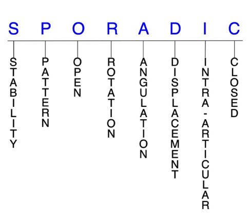
Hand Surgery Resource’s Fracture Description and Characterization Acronym

SPORADIC

S – Stability; P – Pattern; O – Open; R – Rotation; A – Angulation; D – Displacement; I – Intra-articular; C – Closed


S - Stability (stable or unstable)

  • Universally accepted definitions of clinical fracture stability is not well defined in the hand surgery literature.6-8
  • Stable: fracture fragment pattern is generally nondisplaced or minimally displaced. It does not require reduction, and the fracture fragment’s alignment is maintained with simple splinting. However, most definitions define a stable fracture as one that will maintain anatomical alignment after a simple closed reduction and splinting. Some authors add that stable fractures remain aligned, even when adjacent joints are put to a partial range of motion (ROM).
  • Unstable: will not remain anatomically or nearly anatomically aligned after a successful closed reduction and simple splinting. Typically unstable pisiform fractures have significant deformity with comminution, displacement and/or  angulation.
  • Most pisiform fractures are stable and can therefore be treated with immobilization alone.9

P - Pattern

  • Pisiform proximal pole
  • Pisiform distal pole
  • Pisiform body: sagittal, parasagittal, coronal, transverse, and/or comminuted1,10
  • Pisiform fractures are most commonly caused by either direct trauma to the hypothenar eminence—which is frequently seen in sporting activities—or tendon avulsion. Avulsion fractures occur when the flexor carpi ulnaris (FCU) tendon resists forcible hyperextension of the wrist, such as in a  falls on an outstretched hand (FOOSH) injury or when straining to lift a heavy object, which can result in an osteochondral or avulsion fracture to the distal portion of the pisiform. A third mechanism postulated is repetitive trauma causing vascular disruption(AVN), microfractures, and eventually a complete fracture line.11,12  A fall FOOSH injury usually produces a transverse pisiform fracture by a combination of both direct and indirect mechanisms: the pisiform becomes fixed at the pisotriquetral joint when the outstretched hand strikes the ground, and forceful FCU contraction causes a transversely oriented avulsion fracture.1,11

O - Open

  • Open: a wound connects the external environment to the fracture site. The wound provides a pathway for bacteria to reach and infect the fracture site. As a result, there is always a risk for chronic osteomyelitis. Therefore, open fractures of the pisiform require antibiotics with surgical irrigation and wound debridement.6,13,14
  • Open pisiform fractures are exceedingly rare, with the first case in the literature being documented in 2013.3
  • Open fractures of the pisiform may require surgical exploration to determine if articular surfaces are involved. After irrigation and debridement, these wounds are some times left open and further treatment is typically delayed until the wound shows no sign of infection.15,16

R - Rotation

  • Pisiform fracture deformity can be caused by rotation of the distal fragment on the proximal fragment.
  • Degree of malrotation of the fracture fragments can be used to describe the fracture deformity.
  • The pisiform can rotate up to 180° when it is dislocated but rotational deformity is rare.17

A - Angulation (fracture fragments in relationship to one another)

  • Angulation is measured in degrees after identifying the direction of the apex of the angulation.
  • Straight: no angulatory deformity
  • Angulated: bent at the fracture site
  • The small size of the pisiform makes assessing angulation between pisiform fracture fragments difficult.

D - Displacement (Contour)

  • Displaced: disrupted cortical contours
  • Nondisplaced: fracture line defining one or several fracture fragments; however, the external cortical contours are not significantly disrupted
  • Most pisiform fractures are nondisplaced, but they frequently demonstrate intra-articular extension.18
  • The FCU is the wrist’s most powerful muscle, and the pisiform is a sesamoid bone that mechanically enhances its actions. Therefore, the pull of this muscle combined with forced wrist hyperextension associated with a FOOSH cause a significant traction to displace the pisiform proximally usually.  In the presence of ulnar deviation associated with hyperextension of the wrist, the pisiform displaces medially.19

I - Intra-articular involvement

  • Fractures that enter a joint with one or more of their fracture lines.
  • Pisiform fractures can have fragment involvement with the triquetrum at the pisotriquetral joint.
  • Although pisiform fractures are usually nondisplaced, most cases present with intra-articular extension.18
  • If a fracture line enters a joint but does not displace the articular surface of the joint, then it is unlikely that this fracture will predispose to posttraumatic osteoarthritis. If the articular surface is separated or there is a step-off in the articular surface, then the congruity of the joint will be compromised and the risk of posttraumatic osteoarthritis increases significantly. Intra-articular fractures of the pisiform have a particular tendency to develop cartilage degeneration and osteoarthritis, which is why some argue for early mobilization without casting in these cases.20

C - Closed

  • Closed: no associated wounds; the external environment has no connection to the fracture site or any of the fracture fragments.6-8

Pisiform fractures: named fractures, fractures with eponyms and other special fractures

Pisiform dislocation and fracture-dislocation

  • Pisiform dislocations and fracture-dislocations are very rarely reported, but it’s been suggested that many cases are missed and that their actual incidence may be higher.5,21,22
  • Two mechanisms of injury have been described: a direct mechanism by which trauma is applied to the pisiform, and an indirect strong traction on the FCU tendon.  Most pisiform dislocations occur through the latter injury mechanism, while the former is less common and often results in a pisiform fracture instead.21
  • In the more common injury pattern, dislocation occurs with hyperextension traction of the FCU, which can tear the pisohamate and/or pisometacarpal ligaments. This can result from a FOOSH injury that causes significant traction to displace the pisiform proximally. If the pisohamate and pisometacarpal ligaments are damaged, the function of the FCU becomes impaired, with the pisiform retracting proximally.19
  • Pisiform dislocation can occur in isolation or in conjunction with hamate dislocation, distal radius or ulna fractures, and/or other fractures/dislocations.2,5  In most cases, the pisiform dislocates distally and ventrally. There is no clear explanation why this occurs, but it may result from exaggerated physiological translation of the pisiform distally that normally occurs during wrist extension when it hyperextends. It could also result from the FCU tendon failing from being overstretched, or from injuries of ligaments that stabilize the pisotriquetral joint.5,21
  • Common signs and symptoms include ulnar-sided hand pain aggravated by wrist flexion and adduction, swelling, tenderness, loss of hypothenar contour, exaggerated pisiform mobility, and ulnar nerve compression.2,5,21

Imaging

  • Standard radiographic views of the wrist may be difficult to interpret and pisiform injuries may be missed or masked by associated wrist injuries.  Oblique views usually require a comparison image of the contralateral wrist.  In addition to standard radiographs of the wrist, a 30° or 45° oblique supinated view (ball cacher's view) is often needed to visualize the pisotriquetral joint.19,21
  • CT scan is useful for suspected cases and can effectively demonstrate the integrity of the pisiform and identify any associated bony injuries.19,21
  • MRI is also helpful for highlighting any ligamentous or ulnar nerve injuries present.19

Treatment

  • Due to the rarity of pisiform dislocations, an optimal management strategy has not yet been identified.19
  • Most experts recommend a conservative approach for acute dislocations initially, especially if ulnar nerve neurapraxia is present. This consists of immediate closed reduction using wrist flexion and forearm pronation, followed by immobilization with a splint or cast maintaining the reduced position for ~3 weeks.5,19
  • In cases of recurrent dislocation, debilitating chronic pain, or when closed reduction is unsuccessful, surgery is typically needed and may consist of open reduction and internal fixation (ORIF) or pisiform resection or excision.5,19  Removal of the pisiform does not interfere with the normal functioning of the wrist.5
  • When a pisiform dislocation is associated with a distal radius or ulnar fracture, treatment should address both injuries concomitantly.2
  • If the FCU tendon, pisohamate or pisometacarpal ligaments are also damaged, they should be surgically repaired and probably best approached with repair of the FCU tendon possibly with a graft..19,21

Complications

  • Infection
  • Recurrent instability
  • Osteoarthritis
  • Synovitis

Outcomes

  • Outcomes after closed or open reduction and immobilization are mixed, while pisiform resection has been found to offer a more predictable outcome. In cases of delayed diagnosis, resection is also preferred over reduction and is more likely to result in symptomatic relief.19  One reason open reduction leads to poor outcomes may be because it requires dissection, which can lead to more destabilization of the pisiform and damage to its blood supply.21
  • Another study pointed out that good outcomes have been obtained without any treatment or splinting.21

Related Anatomy

  • The pisiform is a small, pea-shaped sesamoid bone located in the most ulnar position of the proximal carpal row, on the volar aspect of the wrist. It consists of a distal pole, a proximal pole, and a body, and it articulates only with the triquetrum—which contributes no stability—on its slightly concave dorsal surface. Its volar surface is round and rough, and serves as an important point of attachment for the flexor retinaculum, the FCU tendon, and the origin of abductor digiti minimi. The pisiform also forms the ulnar wall of Guyon's canal, which is why ulnar nerve symptoms are common in pisiform injuries.5,11,12
  • Ligamentous attachments of the pisiform include the flexor retinaculum, extensor retinaculum, and the pisohamate, pisometacarpal, and pisotriquetral ligaments, which maintain the position of the pisiform upon normal ROM of the wrist.The pisohamate and pisometacarpal ligaments, together with the ulnar part of the pisotriquetral ligament, are the primary stabilizers of the pisiform. There is also a superficial fibrous band that extends from the pisiform to the hook of hamate.1,2,11,12
  • The pisiform is the only carpal bone that receives direct tendinous insertion, and the tendons associated with it include the FCUand the abductor digiti minimi. The pisiform is not completely embedded in the FCU, but it attaches to the tendon’s anterior surface, and it also forms an origin of the abductor digiti minimi. The FCU tendon continues distally to the pisohamate and pisometacarpal ligaments, which insert upon the hook of hamate and the bases of the ring and little metacarpals, respectively.2,5,12
  • Carpal tunnel release can cause post-operative painful pisiform subluxation.

Incidence and Related injuries/conditions

  • Fractures of the carpal bones have been found to account for 8-18% of all hand fractures23,24and ~6% of fractures overall.25
  • Fractures of the proximal carpals are more common than the distal carpals, and the most commonly fractured carpal bone is the scaphoid, which represents 58-89% of all carpal fractures.23,24,26,27
  • Fractures of the other 7 carpals are very rare and only account for ~1.1% of all fractures. The triquetrum ranks highest of these bones, while fractures of the remaining carpals are even less common and vary in incidence.28-30
  • The incidence of pisiform fractures has been estimated at between 0.2-2% of all carpal fractures.11,24,27,31
  • About 50% of pisiform fractures occur in isolation, while the other 50% are associated with other wrist injuries, such as a perilunate dislocation, distal radius fracture, or additional carpal fracture.1,11

Work-up Options

  • Routine X-rays
    • Pisiform fractures are difficult to recognize on routine radiographic views because of the fracture’s orientation, improper wrist positioning, superimposition of adjacent bones, or an inadequate number of projections. Nonetheless, it is advisable to use these films as the first step in evaluating possible pisiform fracture because they are readily available and inexpensive.
    • A posteroanterior (PA) view will demonstrate transverse and sagittal fractures, while a lateral projection may show coronal fractures; however the pisiform may be obscured by overlapping other bones on both the lateral and standard oblique views.11
  • Special X-ray views
    • A 30° or 45° supinated oblique view in slight extension is helpful for demonstrating fractures in the coronal plane and also shows the pisotriquetral joint.10,11
    • A carpal tunnel view is also beneficial for showing coronal fractures, but this view can be difficult to obtain in the acute setting, as dorsiflexion is limited by pain.10-12
    • A clenched fist PA view with the wrist held in ulnar deviation may also assist the diagnosis.2
  • CT scan
    • Typically recommended if suspicion of fracture is high, other exams are negative, and the patient remains symptomatic, although some authors recommend systematic use of CT scan for all wrist injuries.3,11
    • CT scans can provide useful additional information about fracture morphology and displacement, especially if they are not in the scan plane.2,11
  • MRI
    • Useful for confirming a pisiform fracture diagnosis, especially if radiographs were negative and suspicion remains. May show bone marrow edema within the pisiform, which can indicate a fracture and associated soft tissue injuries.4
  • Bone scan
ICD-10 Codes

PISIFORM FRACTURE

Diagnostic Guide Name

PISIFORM FRACTURE

ICD 10 Diagnosis, Single Code, Left Code, Right Code and Bilateral Code

DIAGNOSIS SINGLE CODE ONLY LEFT RIGHT BILATERAL (If Available)
FRACTURE CARPAL BONE - PISIFORM        
- DISPLACED   S62.162_ S62.161_  
- NONDISPLACED   S62.165_ S62.164_  

Instructions (ICD 10 CM 2020, U.S. Version)

THE APPROPRIATE SEVENTH CHARACTER IS TO BE ADDED TO EACH CODE FROM CATEGORY S62
  Closed Fractures Open Type I or II or Other Open Type IIIA, IIIB, or IIIC
Initial Encounter A B C
Subsequent Routine Healing D E F
Subsequent Delayed Healing G H J
Subsequent Nonunion K M N
Subsequent Malunion P Q R
Sequela S S S

ICD-10 Reference

Reproduced from the International statistical classification of diseases and related health problems, 10th revision, Fifth edition, 2016. Geneva, World Health Organization, 2016 https://apps.who.int/iris/handle/10665/246208

Clinical Presentation Photos and Related Diagrams
Pisiform Fracture Diagrams
  • Non-displaced pisiform fracture (arrow).
    Non-displaced pisiform fracture (arrow).
  • Displaced pisiform fracture (arrow)
    Displaced pisiform fracture (arrow)
Symptoms
History of trauma to the ulnar palmar wrist
Fracture pain, swelling, and tenderness are typically concentrated over the proximal hypothenar eminence.12
Swelling and ecchymosis
Fracture deformity and crepitus are very rare
Ulnar nerve compression symptoms
Loss of grip strength
Typical History

A typical patient is a 34-year-old right-handed woman who is an avid mountain biker. During a recent ride, she failed to see a divot on the bike trail and rode her front wheel directly into it, which caused the bike to flip forward and sent her over the handlebars. She landed a few feet in front of the divot with her hands outstretched, wrists extended, and forearms pronated and adducted.  Her left wrist absorbing the brunt of the impact and the force created by her body weight accelerating through the air during the fall. The force fractured the pisiform and triquetrum of her left wrist, and resulted in severe pain and inflammation, which led her to seek out immediate treatment at the emergency department.

Treatment Options
Treatment Goals
  • When treating closed pisiform fractures, the treating surgeon has 4 basic goals:6,14
    1. A pisiform with a normal appearance. The X-ray may not need to be perfect, but the pisiform should have no obvious deformity (ie, the pisiform looks normal!)
    2. Avoid stiffness by maintaining a normal functional range of motion (ROM)  (ie, the wrist works!)
    3. The pisiform is not painful (ie, the pisiform does not hurt!)
    4. Congruent joint surface with none-to-minimal joint surface irregularities (ie, the pisotriquetral joint does not develop early posttraumatic arthritis!)
    5. Fracture care for open injuries should also minimize the risk for infection and osteomyelitis.
Conservative
  • There is no consensus on the optimal treatment approach for pisiform fractures on account of their rare occurrence—especially acutely diagnosed cases—but because most of these injuries are stable and nondisplaced, most experts seem to agree that conservative treatment is usually sufficient.1,9,11
  • Conservative treatment is particularly indicated for avulsion fractures, parasagittal fractures, and nondisplaced transverse intra-articular fractures of the pisiform.10,32  Casting typically consists of immobilization with 30° of wrist flexion and ulnar deviation using a spica cast or aluminum, plaster, fiberglass, or custom splint for 3-6 weeks. Bony or asymptomatic fibrous union usually occurs within 4-6 weeks.5,8,12,16
  • These injuries should be subsequently reexamined and reimaged to confirm healing.32  Some authors have even made an argument for early mobilization without casting for intra-articular pisiform fractures, since these injuries have a tendency to develop cartilage degeneration and osteoarthritis.20
Operative
  • Surgical treatment of pisiform fractures must always be an individualized therapeutic decision. However, surgical pisiform fracture care is most frequently recommended when:
    1. Closed reduction fails or the simple splint or cast immobilization does not maintain the reduction. For these irreducible or unstable fractures, operative treatment is recommended to achieve the 4 treatment goals of fracture care.
    2. There is a significantly displaced pisiform fracture fragment involving the pisotriquetral joint. Surgical fracture care may be required in these cases.
    3. Open pisiform fractures require surgical care in the form of irrigation and debridement to prevent chronic infection.
  • For pisiform fractures that are displaced and/or comminuted, and cases that fail conservative treatment and/or develop complications—like osteoarthritis, refractory pain, or ulnar neuropathy and surgery is typically needed.  The most commonly utilized surgical procedure and treatment-of-choice for these cases is pisiform excision (pisiformectomy), which is performed either in an early or delayed fashion.9,20,32
  • Some authors have recommended a volar approach on the radial side of the FCU tendon, which allows clear visualization of the ulnar nerve and decompression of the pisohamate ligament. An ulnar approach is not advocated because of the hazard of the nearby dorsal cutaneous branch of the ulnar nerve and potential for painful neuroma formation.2
  • Following excision, the use of padded gloves may alleviate wound tenderness and enable early return to sporting activity.2  Another useful surgical approach is splitting the FCU tendon.
  • Cases with symptomatic posttraumatic osteoarthritis or nonunion may also require FCU tendon reconstruction.1
  • There are no reports of internal fixation for pisiform fractures in the literature, and some have therefore concluded that this intervention does not have a role in the management process.2

Post-treatment Management

  • The care and precautions related to immobilization devices for the pisiform fracture must be carefully reviewed with the patient. Patients should be educated regarding care and precautions. Patients should know that pain, especially increasing pain, numbness, tingling, skin irritation, splint loosening, or excessive splint tightness are red flags and should be reported to the surgeon or his team.
  • Pain should be managed with properly fitting splints, reassurance, elevation, ice in the initial post-fracture period, and mild pain medications. Patients should be encouraged to discontinue pain medication as soon as possible. Opioid use should be kept to a minimum.
  • Nondisplaced pisiform fractures can be expected to heal reliably within 4-6 weeks of immobilization.2
  • If an infection does occur in association with an open fracture then management should focus on eradicating sepsis with thorough debridement, appropriate antibiotics (eg, cephalosporin, penicillin), and pisiform excision.16
  • Patients should be instructed to carefully exercise all joints in the injured hand, wrist, and arm that do not require immobilization.  Patients usually can exercise on their own; however, signs of generalized hand or wrist stiffness are indications for referral to hand therapy (PT or OT).
Treatment Photos and Diagrams
Pisiform Fracture Treatment
  • Short arm cast treatment for an acute non-displaced pisiform fracture.
    Short arm cast treatment for an acute non-displaced pisiform fracture.
  • Wrist brace treatment for an non-acute pisiform fracture without displacement
    Wrist brace treatment for an non-acute pisiform fracture without displacement
  • Excision of the pisiform for pisotriquetral osteoarthritis through the proximal part of an extended carpal tunnel incision. Pisiform (P); Neurovascular bundle (NVB); Flexor car ulnas (FCU)
    Excision of the pisiform for pisotriquetral osteoarthritis through the proximal part of an extended carpal tunnel incision. Pisiform (P); Neurovascular bundle (NVB); Flexor car ulnas (FCU)
  • Excision of the pisiform. The FCU has been splitted and the attachments to the volar pisiform are being removed sharply. Pisiform (P); Flexor Carpi Ulnas (FCU)
    Excision of the pisiform. The FCU has been splitted and the attachments to the volar pisiform are being removed sharply. Pisiform (P); Flexor Carpi Ulnas (FCU)
  • Excision of the pisiform. The pisotriqutral ligaments as the final step in pisiform excision.  Pisiform (P); Flexor Carpi Ulnaris (FCU)
    Excision of the pisiform. The pisotriqutral ligaments as the final step in pisiform excision. Pisiform (P); Flexor Carpi Ulnaris (FCU)
  • Excision of the pisiform. Pisiform completely removed (P). The neurovasciular bundle (NVB) i.e. ulnar artery and nerve intact. Flexor Carpi Ulnaris (FCU) split to be sutured before closure.
    Excision of the pisiform. Pisiform completely removed (P). The neurovasciular bundle (NVB) i.e. ulnar artery and nerve intact. Flexor Carpi Ulnaris (FCU) split to be sutured before closure.
CPT Codes for Treatment Options

Per an agreement between Hand Surgery Resource, LLC and the American Medical Association (AMA) users are required to accept the following End User Point and Click Agreement in order to view CPT content on this website.  Please read and then click "Accept" at the bottom to indicate your acceptance of the agreement.

End User Point and Click Agreement

CPT codes, descriptions and other data only are copyright 2019 American Medical Association (AMA). All rights reserved. CPT is a registered trademark of the AMA. The AMA reserves all rights to approve any license with any Federal agency.

You, as an individual, are authorized to use CPT only as contained in Hand Surgery Resource solely for your own personal information and only within the United States for non-commercial, educational use for the purpose of education relating to the fundamental principles of hand surgery and the common diseases, disorders and injuries affecting the human hand. You agree to take all necessary steps to ensure your compliance with the terms of this agreement.

Any use not authorized herein is prohibited, including by way of illustration and not by way of limitation, making copies of CPT for resale and/or license, transferring copies of CPT to any party not bound by this agreement, creating any modified or derivative work of CPT, or making any commercial use of CPT. License to use CPT for any use not authorized herein must be obtained through the AMA, CPT Intellectual Property Services, 330 N. Wabash Avenue, Chicago, IL 60611. Applications are available at the AMA Web site, http://www.ama-assn.org/go/cpt.

Common Procedure Name
Excision trapezium, pisiform, scaphoid of hook of hamate
CPT Description
Carpectomy; one bone
CPT Code Number
25210
CPT Code References

The American Medical Association (AMA) and Hand Surgery Resource, LLC have entered into a royalty free agreement which allows Hand Surgery Resource to provide our users with 75 commonly used hand surgery related CPT Codes for educational promises. For procedures associated with this Diagnostic Guide the CPT Codes are provided above. Reference materials for these codes is provided below. If the CPT Codes for the for the procedures associated with this Diagnostic Guide are not listed, then Hand Surgery Resource recommends using the references below to identify the proper CPT Codes.

 CPT QuickRef App.  For Apple devices: App Store. For Android devices: Google Play

 CPT 2021 Professional Edition: Spiralbound

Complications
  • Complications are generally rare in pisiform fractures, but may include nonunion, painful union, malunion, ulnar nerve compression, and chondromalacia, posttraumatic osteoarthritis, or subluxation of the pisotriquetral joint.2,11,12,33
  • Although nonunion, malunion, and painful union are uncommon, these complications are often disabling when they do occur, and may cause chronic pain, weakness, or impaired wrist ROM.11,33
  • Ulnar nerve compression symptoms may also result from damage to the ulnar nerve in Guyon's canal, and these cases usually require decompression of the ulnar nerve.33
  • Pisotriquetral chondromalacia, subluxation, and osteoarthritis are possible complications if the articular surface is poorly aligned at the pisotriquetral joint.12
Outcomes
  • Both conservative and surgical methods have been found to elicit positive outcomes after pisiform fractures, and neither approach has been clearly identified as superior to the other.1,2,20,33,34
  • Pisiform fractures that have a transverse rather than a longitudinal orientation tend to functionally disrupt the FCU function and generally have a more guarded prognosis.1
  • In cases of nonunion, malunion, or a displaced acute pisiform fractures, pisiform excision has been found to result in a predictably satisfactory outcomes and an early return to function.2
  • In one study, 67 patients had pain and dysfunction of the pisotriquetral joint—42 of which had a previous history of fracture—and all were treated surgically with pisiform excision.  At surgery, osteoarthritic changes were seen in 20 joints and pisotriquetral chondromalacia in 29 patients.  Complete symptomatic relief was obtained in 65 wrists without loss of wrist strength or ROM, and the authors also noted a lack of postoperative problems with the FCU tendon.34
  • In another study, 21 of 33 patients over 10 years were treated with pisiform excision for pisiform area pain after failing conservative treatment, with 5 having pisiform fracture and 6 with pisotriquetral osteoarthritis.   Nineteen of these patients (90%) had relief of their symptoms and all regained their preoperative wrist ROM.33
Key Educational Points
  • Underlying pathological conditions such as bone tumors—like enchodromas—and osteoporosis should be expected in fractures that occur from trivial trauma.
  • The functional needs of each patient must be considered when recommending treatment for pisiform fractures.
  • Although non-scaphoid carpal bones like the pisiform have received considerably less attention than the scaphoid, these injuries can still produce morbidity that is disproportionate to their incidence for 2 primary reasons:  (1) - These fractures may have a subtle clinical and radiographic presentation and are easily overlooked or misdiagnosed, in some cases as wrist sprains. This can lead to suboptimal management and poor long-term outcomes with significant wrist disability.  (2) - These fractures are often harbingers of significant ligamentous disruption or associated carpal fractures, and failure to recognize a more global injury pattern can result in undertreatment and permanent wrist dysfunction.1
  • Although pisiform fractures are uncommon, their incidence seems to be increasing due to the popularity of golf and other racquet sports.2
  • It’s important to distinguish pisiform fractures from other causes of hypothenar pain and ulnar sided wrist pain like hamate fracture, FCU tendinitis, carpal ligamentous disruptions, and pisotriquetral chondromalacia and osteoarthritis.11
References

New and Cited Articles

  1. Shah MA, Viegas SF. Fractures of the carpal bone excluding the scaphoid. J Hand Surg Am 2002; 2(3): 129-140
  2. O'Shea K, Weiland AJ. Fractures of the hamate and pisiform bones. Hand Clin2012;28(3):287-300. PMID: 22883867
  3. Agathangelidis F, Boutsiadis A, Ditsios K. Open pisiform fracture: excision or internal fixation?BMJ Case Rep2013;2013. pii: bcr2012007621. PMID: 23307459
  4. Boyce SH, Harden JL. Pisiform fracture: an uncommon wrist injury. BMJ Case Rep2010;2010:2132. PMID: 22736214
  5. Muñiz AE. Unusual wrist pain: pisiform dislocation and fracture. Am J Emerg Med1999;17(1):78-9. PMID: 9928708
  6. Cheah AE, Yao J. Hand Fractures: Indications, the Tried and True and New Innovations.J Hand Surg Am 2016;41:712-22. PMID: 27113910
  7. Nesbitt KS, Failla JM, Les C. Assessment of instability factors in adult distal radius fractures. J Hand Surg Am 2004;29:1128-38.PMID: 15576227
  8. Walenkamp MM, Vos LM, Strackee SD, Goslings JC, Schep NW. The Unstable Distal Radius Fracture-How Do We Define It? A Systematic Review. J Wrist Surg 2015;4:307-16. PMID: 26649263
  9. Husband JB. Hook of hamate and pisiform fractures in basketball and hockey players. Hand Clin2012;28(3):303. PMID: 22883869
  10. Suh N, Ek ET, Wolfe SW. Carpal fractures. J Hand Surg Am2014;39(4):785-91. PMID: 24679911
  11. Fleege MA, Jebson PJ, Renfrew DL, et al. Pisiform fractures. Skeletal Radiol1991;20(3):169-72. PMID: 2057787
  12. Lacey JD, Hodge JC. Pisiform and hamulus fractures: easily missed wrist fractures diagnosed on a reverse oblique radiograph. J Emerg Med1998;16(3):445-52. PMID: 9610976
  13. Ketonis C, Dwyer J, Ilyas AM. Timing of Debridement and Infection Rates in Open Fractures of the Hand: A Systematic Review. Hand (N Y) 2017;12:119-26. PMID: 28344521
  14. Meals C, Meals R. Hand fractures: a review of current treatment strategies. J Hand Surg Am 2013;38:1021-31. PMID: 23618458
  15. Day CS. Fractures of the Metacarpals and Phalanges. In: Green DP, ed. Green's Operative Hand Surgery. Seventh ed. Philadelphia: Elsevier; 2016, pp. 231-77.
  16. Weinstein LP, Hanel DP. Metacarpal fractures.J Hand Surg Am 2002; 2(4):168–180.
  17. Minami M, Yamazaki J, Ishii S. Isolated dislocation of the pisiform: a case report and review of the literature. J Hand Surg Am1984;9A(1):125-7. PMID: 6693731
  18. Zimmerman NB, Mass DP. A pisiform fracture. Orthopedics1987;10(5):817-20. PMID: 3588426
  19. Goriainov V, Bayne G, Warwick DJ. Traumatic dislocation of the pisiform: a case report. J Orthop Surg (Hong Kong)2010;18(3):389-90. PMID: 21187560
  20. Mattelaer B, De Smet L. Bilateral pisiform fractures. J Hand Surg Eur Vol2010;35(3):242-3. PMID: 20200081
  21. Saleh WR, Yajima H, Nakanishi A. Conservative treatment of the isolated dislocation of the pisiform bone. J Plast Surg Hand Surg2014;48(4):283-4. PMID: 23834301
  22. Gainor BJ. Simultaneous dislocation of the hamate and pisiform: a case report. J Hand Surg Am1985;10(1):88-90. PMID: 3968411
  23. van Onselen EB, Karim RB, Hage JJ, Ritt MJ. Prevalence and distribution of hand fractures. J Hand Surg Br2003;28(5):491-5. PMID: 12954264
  24. Hove LM. Fractures of the hand. Distribution and relative incidence. Scand J Plast Reconstr Surg Hand Surg1993;27(4):317-9. PMID: 8159947
  25. Dobyns JH, Beckenbaugh RD, Bryan RS, et al. Fractures of the hand and wrist. In: Flynn JE, editor. Hand surgery. Third ed. Philadelphia: Lippincott Williams & Wilkins; 1982.
  26. Rhemrev SJ, Ootes D, Beeres FJ, et al. Current methods of diagnosis and treatment of scaphoid fractures. Int J Emerg Med2011;4:4. PMID: 21408000
  27. Hey HW, Chong AK, Murphy D. Prevalence of carpal fracture in Singapore. J Hand Surg Am2011;36(2):278-83. PMID: 21276891
  28. Larsen CF, Brøndum V, Skov O. Epidemiology of scaphoid fractures in Odense, Denmark. Acta Orthop Scand1992;63(2):216-8. PMID: 1590062
  29. Höcker K, Menschik A. Chip fractures of the triquetrum. Mechanism, classification and results. J Hand Surg Br1994;19(5):584-8. PMID: 7822914
  30. Garcia-Elias M.Dorsal fractures of the triquetrum-avulsion or compression fractures? J Hand Surg Am1987;12(2):266-8. PMID: 3559084
  31. Geissler WB. Carpal fractures in athletes. Clin Sports Med2001;20(1):167-88. PMID: 11227704
  32. Hulsopple C, Deluca J, Jonas C. Treatment of Acute Carpal Bone Fractures. Curr Sports Med Rep2017;16(5):330-335. PMID: 28902755
  33. Palmieri TJ. Pisiform area pain treatment by pisiform excision. J Hand Surg Am1982;7(5):477-80. PMID: 7130657
  34. Carroll RE, Coyle MP Jr. Dysfunction of the pisotriquetral joint: treatment by excision of the pisiform. J Hand Surg Am1985;10(5):703-7. PMID: 4045152
  35. Minami M, Yamazaki J, Ishii S. Isolated dislocation of the pisiform: a case report and review of the literature. J Hand Surg Am1984;9A(1):125-7. PMID: 6693731
  36. Matsumoto T, Tsunoda M, Yamaguchi S, et al. Traumatic dislocation of the hamate and pisiform: a case report and review of the literature. J Orthop Trauma2005;19(4):282-5. PMID: 15795579

Classic

  1. Deane RB. Simple Fracture of the Pisiform Bone.Ann Surg1911;54(2):229. PMID: 17862721